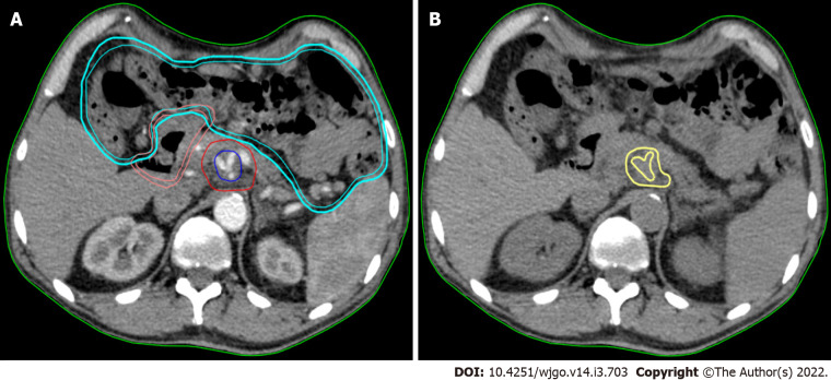
Figure 1.
Texture analysis performed using contrast-free simulation computed tomography imaging. A: Target volumes delineation in a stereotactic ablative radiation therapy case. The high-dose planning target volume (blue) encompasses the tumour-vessel interface (TVI, encasement of celiac axis) inside the tumour planning target volume (PTVt, red). The prescription dose is 30 Gy and 50 Gy in 5 fractions, with simultaneous integrated boost, to PTVt and TVI, respectively. The following organ at risk are shown: duodenum (pink) and bowel (cyan); B: The gross tumour volume (GTV) of the pancreatic lesion without vessels (yellow) is shown in the same axial computed tomography (CT)-simulation image without contrast. This is the final CT used for analysis.

