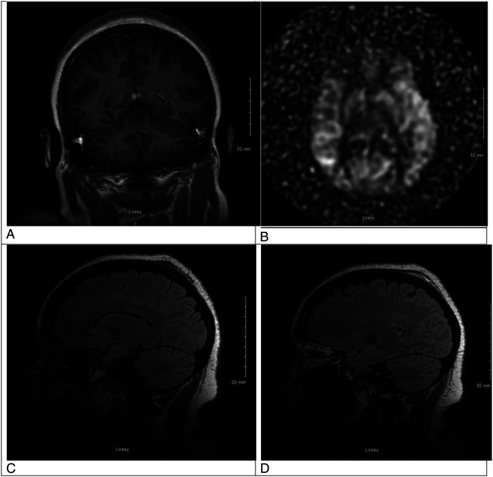
Figure 2.
A. This is an MRI brain coronal T1 post contrast view showing resolution of the previously contrast enhancing lesion in the left hemisphere. B. This is an MRI brain arterial spin labeling sequence showing resolution of the previous relative hyperperfusion of the left hemisphere. C. This is an MRI brain sagittal T2 Flair image without contrast showing resolution of the previous frontal lobe edema. D. This is an MRI brain sagittal T2 Flair with contrast showing resolution of enhancement the prior left occipital lobe lesion.

