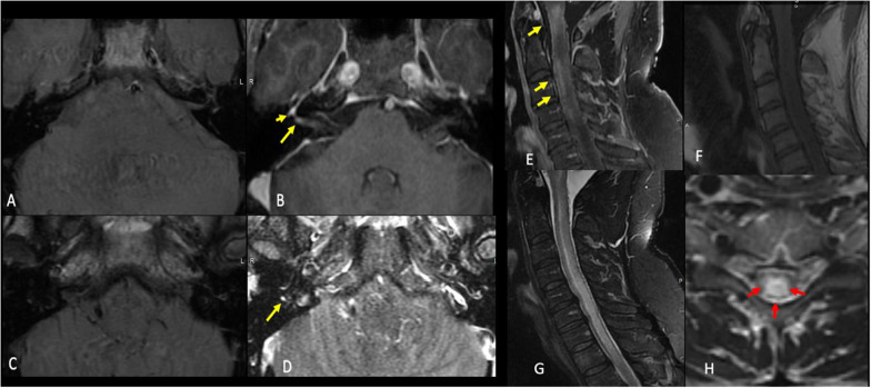
Fig. 5.
Neuroimaging features after COVID-19 vaccination. Case 1: A 46-year-old-man who presented with a rapid onset right-sided facial weakness after having COVID-19 vaccine. Axial T1 pre (a and c) and T1 postcontrast fat sat (b and d) demonstrate abnormal enhancement of the right facial nerve within the lateral right IAC (long yellow arrow in b) as well as asymmetric enhancement of the right geniculate ganglion (short yellow arrow in b) and tympanic portion of the facial nerve (yellow arrow in d). Findings are consistent with Bell's palsy. Case 2: A 51-year-old-man who presented with a sudden upper and lower limb weakness after having COVID-19 vaccine. Sagittal T1 postcontrast (e) T1 pre (f), STIR (g), and axial T2W (h) images demonstrate extensive T2 signal hyperintensity of the central cervical cord (red arrows) with patchy areas of enhancement at the levels of c1, c2, c3 and c4 (yellow arrows). There was no associated restricted diffusion. Findings were consistent with transverse myelitis

