Fig. 3.
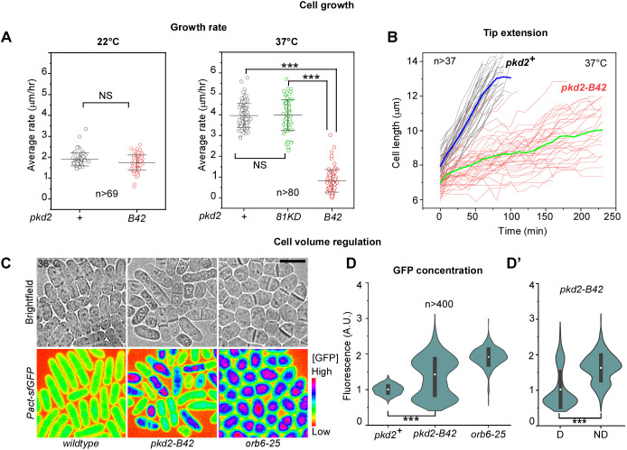
Pkd2p is required for cell size expansion during interphase growth. (A) Dot plots of the average tip growth rate of the wild-type (pkd2+) and the pkd2 mutant cells at 22°C (left) and 37°C (right). Line and error bars show the mean±s.d. (B) Time courses of the length extension of wild-type and pkd2-B42 cells at 37°C. Thick lines (blue, wild type; green, pkd2-B42) represent the mean time courses, n>37. (C) Brightfield (top) and fluorescence micrographs (bottom, spectrum-colored) of wild-type (left), pkd2-B42 (middle) and orb6-25 (right) cells at 36°C. All strains constitutively expressed super-folder GFP under control of the Pact promoter (Pact-sfGFP). The fluorescence intensity is inversely correlated with the volume expansion rate. Scale bar:10 µm. (D) Violin plots of intracellular GFP concentration in the indicated strains. (D’) The intracellular GFP concentration of the deflated (D) and non-deflated (ND) pkd2-B42 cells, n>400. Black bars indicate s.d. and white dots mark the mean. The data were pooled from at least two independent biological repeats. A.U., arbitrary units. ***P<0.001; NS, not significant (two-tailed unpaired Student's t-test).

