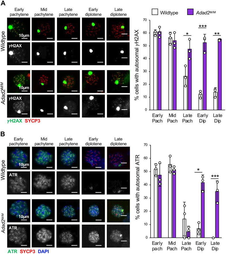
Fig. 4.
Loss of ADAD2 results in abnormal autosomal γH2AX and ATR late in meiosis. (A) Quantification of autosomal γH2AX signal by spermatocyte stage in 30 dpp wild-type and Adad2M/M germ cells (H2AX, green; SYCP3, red) showing mutant-specific increase of cells with autosomal γH2AX in late pachytene through diplotene. (B) Quantification of autosomal ATR signal by spermatocyte stage in 30 dpp wild-type and Adad2M/M germ cells (three samples per genotype; ATR, green; SYCP3, red; DAPI, blue) showing increased frequency of autosomal ATR in mutant spermatocytes late in meiosis. Images are representative of localization pattern observed in wild type versus mutant. Data are mean±s.d. Dots represent frequencies within individuals. Significance was calculated using an unpaired, two-tailed Student‘s t-test (*P<0.05, **P<0.001, ***P<0.0001).

