Fig. 3.
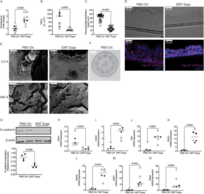
Comparative analysis of epithleial plasticity of pEMT in primary non-diseased human bronchial epithelia. (A) NHBE cells undergoing pEMT induced by treatment with 2× EMT inducer supplement (EMT supp) display increased FITC–dextran permeability compared to control PBS-treated (PBS Ctrl) cells. (B) NHBE cells undergoing pEMT display decreased TEER. (C) NHBE cells undergoing pEMT have a lower height of pseudostratified epithelium. (D) There is an expected loss of apical-basal polarity in the cells undergoing EMT as determined by immunofluorescence (basolateral, Na+/K+-ATPase, magenta). Taken with a 40× oil objective. Scale bars: 25 µm. (E) There is a significant loss of specialized apical structures in cells undergoing pEMT such as cilia, as assessed using scanning electron microscopy. Images taken at 2.5× and 800×. Scale bars: 2 µm, 10 µm. (F) Using transmission electron microscopy, cilia were identified in NHBE treated with PBS Ctrl (top panel); however, there were no cilia evident to capture in the EMT sample, highlighting the loss of specialized apical structures. Images in D–F representative of three donors per group (two inserts per donor). (G) Representative blots (top panel) and quantification (bottom panel) showing decreased E-cadherin expression in cells treated with EMT Supp compared to those treated with PBS Ctrl as detected by western blotting. β-actin levels are shown as a loading control. Blot representative of four inserts from one donor. (H–N) Basal mRNA expression of the epithelial marker CDH1 is lower in pEMT-induced NHBE cells (H), and levels of mesenchymal markers (CDH2, VIM, SNAI1, SNAI2, ZEB1 and ZEB2) relative to GAPDH are increased (I–N) with treatment with EMT Supp were compared to PBS Ctrl, as analyzed by qPCR. Data is generated from three donors (two to four inserts per donor). Data is expressed with median bars. Statistics determined by Mann–Whitney test, with P<0.05 considered statistically significant.

