Fig. 7.

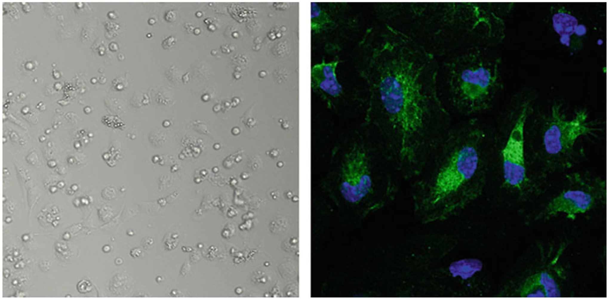
Representative image of the Kupffer cells. (Left) Bright-field image of the Kupffer cells in culture the day after plating. (Right) Kupffer cells stained with F4/80 antibody (green) and DAPI (blue)

Representative image of the Kupffer cells. (Left) Bright-field image of the Kupffer cells in culture the day after plating. (Right) Kupffer cells stained with F4/80 antibody (green) and DAPI (blue)