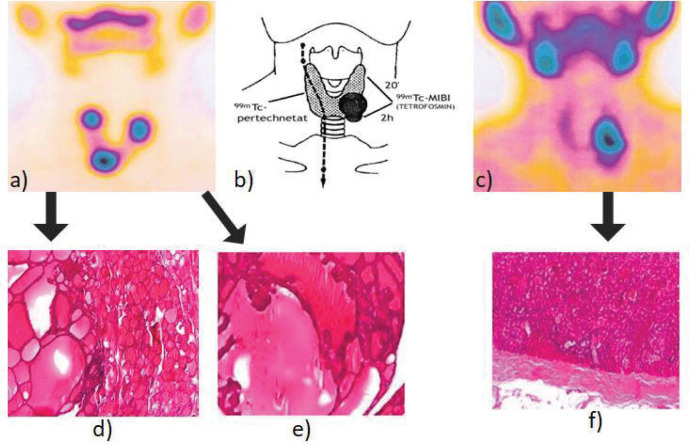
Figure 1.
Thyroid/parathyroid images with two radiotracers, 99mTcO4- (a) and 99mTc sestaMIBI (early scan - at 20 minutes after iv administration) (c), showed comparatively, in addition to the parathyroid scintigraphy principle (b). The nodular formation described in ultrasounds in the left lobe area has an intense 99mTc-sestaMIBI uptake, discordant to the correspondent 99mTcO4-uptake, that presents a totally different nodular uptake pattern. Histopathology confirmed the image findings: colloidal goiter with macrofollicular adenomatous areas and lymphocytic thyroiditis, HE, x4 (d), alternated with macrofollicular adenomatous area with hyperfunctional pseudopapilla, HE, x4 (e) and parathyroid adenoma with oxyphil cells, trabecular and acinar architecture, HE, x4 (f).

