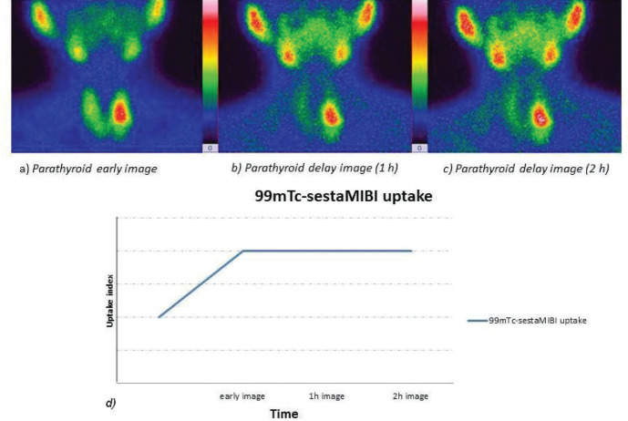
Figure 2.
The high 99mTc-sestaMIBI uptake in the projection area of the 1/3 region of the left thyroid lobe, in early images, persists with approximately the same intensity in both 1h and 2h delayed images (quantified by the uptake index - higher values for delayed images, in the graph), suggesting a parathyroid adenoma and less probably a malignant substrate.

