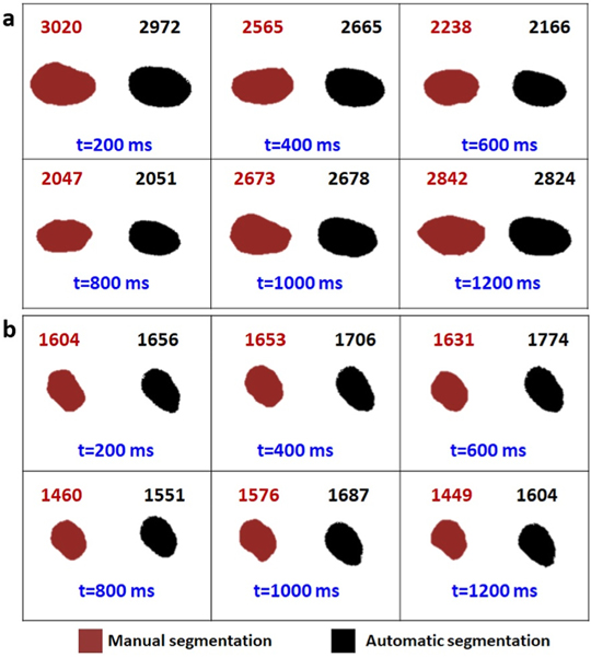
Fig. 6. Validation of U-net image segmentation framework.
The sequential frames from a wild type zebrafish recorded video with fps of 5 are extracted. The respective ventricle mask of each frame is shown in each panel via manual and automatic segmentation. The area of each ventricle is measured and written above its own box. Considering the fps of the videos and the average heart rate of the zebrafish 6 consecutive frames have been shown in this figure to ensure having at least one full cycle.

