
FIG. 1.
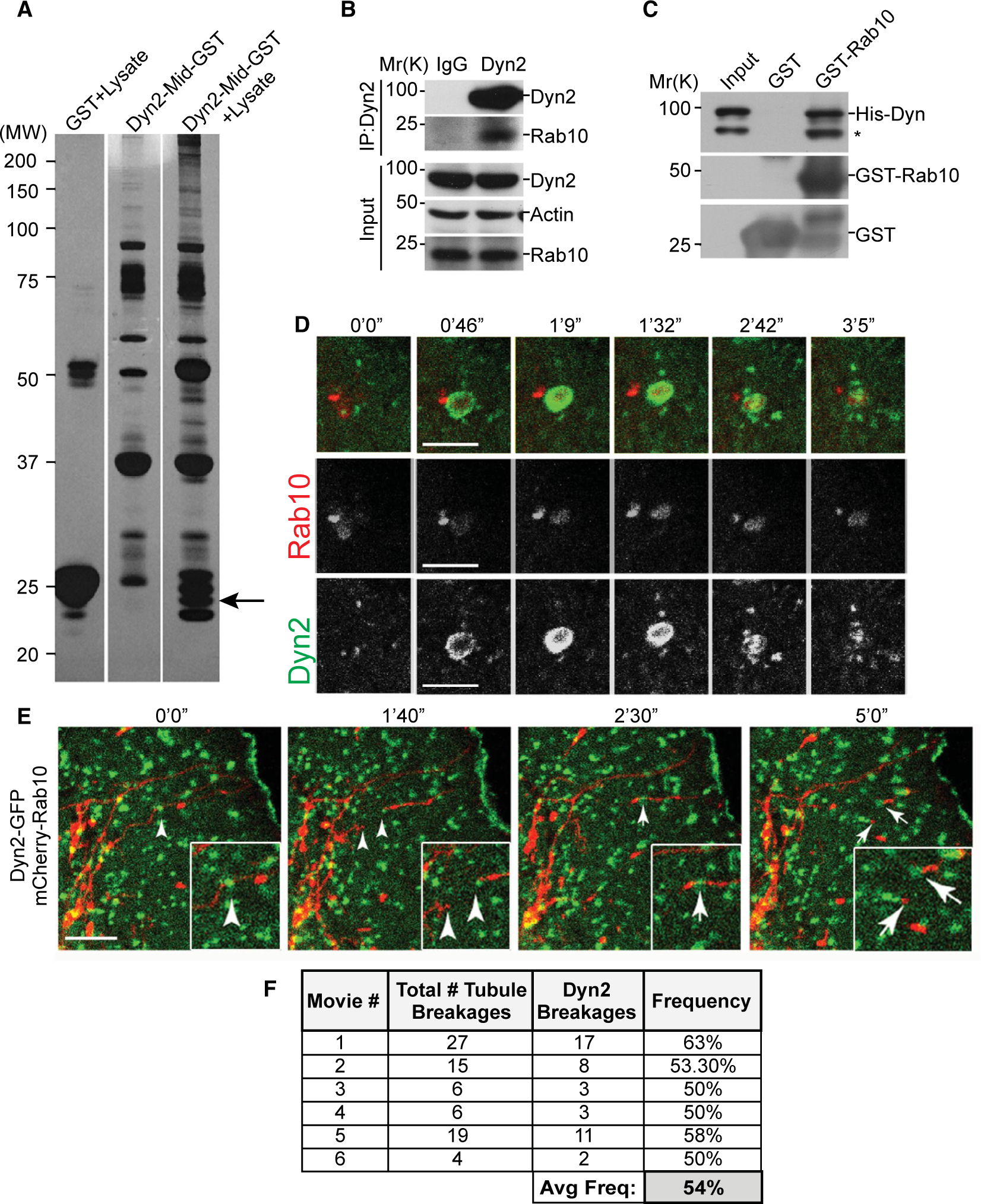
Rab10 and dynamin proteins interact in vitro and in living cells. (A) GST pull-down screen for proteins that bind to the middle domain of Dyn2 (Dyn2-Mid). Putative binding partners isolated from rat brain homogenate visualized by silver stain. (B) Immunoprecipitation of Rab10 by a Dyn2 antibody from Clone9 cell lysates. (C) Immunoblottings from the in vitro GST pull-down experiment using GST-Rab10 and Dyn2-His to examine direct binding. (D) Live-cell microscopy of 24-hour low-serum (0.1% FBS) starved Hep3B hepatoma cells cotransfected with Dyn2-GFP and mCherry-Rab10. (E) Time-lapse stills of a live Hep3B cell following 24-hour low-serum starvation, showing the recruitment of GFP-Dyn2 to a scission point of an mCherry-Rab10–coated membrane tubule. (F) Table displaying number and frequency of mCherry-Rab10 tubule scission events occurring with identifiable Dyn2-GFP puncta at a site of tubule breakage. Micron bars denote 5 μm. Abbreviations: Avg Freq, average frequency; His-Dyn, His-tagged dynamin; IgG, immunoglobulin G; IP, immunoprecipitation; MW, molecular weight.