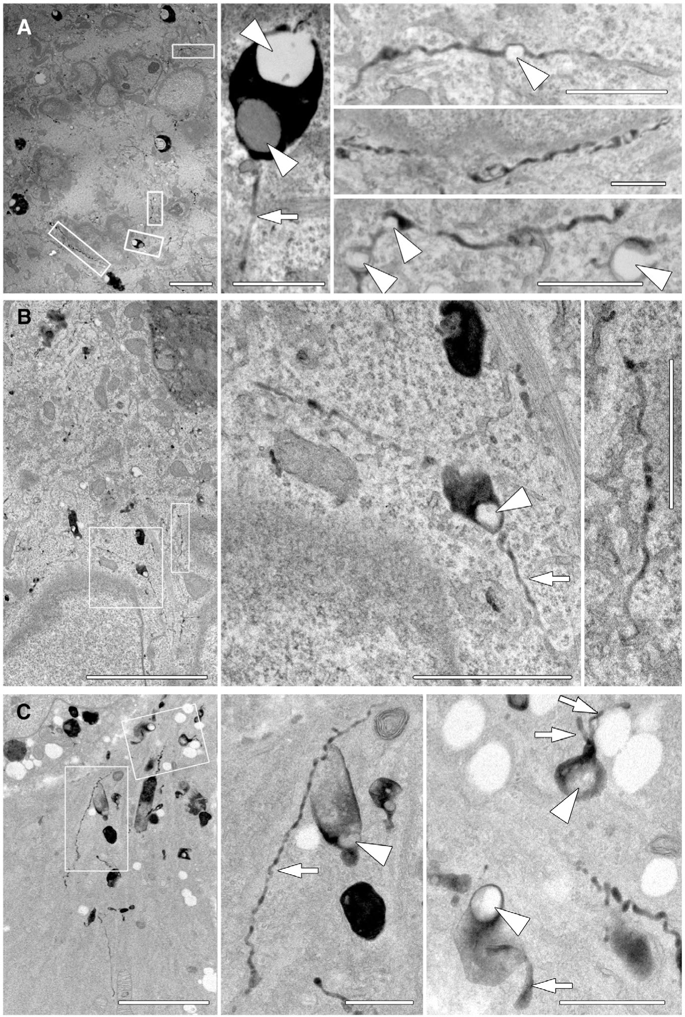
FIG. 5.

EM of thin HRP-positive tubules extending from lipid droplets in cells with reduced Dyn2 levels. Hep3B cells were treated with Dyn2 siRNA for 3 days, then serum starved for 24 hours; cells were then incubated with HRP for 90 minutes, chased 2 hours, and processed for EM and development of the HRP reaction product. (A-C) Hep3B cells that were treated with siRNA targeting endogenous Dyn2. Enlarged panels of boxed areas from low-magnification images show the presence of HRP-positive membranes that extend through the cytoplasm for significant distances. These tubules are not observed in control-treated cells and can encase exceptionally small LDs that would not be resolved by light microscopy. Arrows indicate HRP positive tubules and arrow heads indicate LDs. Micron bars in A-C = 5 μm; all increased magnifications of boxed regions = 1 μm.
