Figure 4.

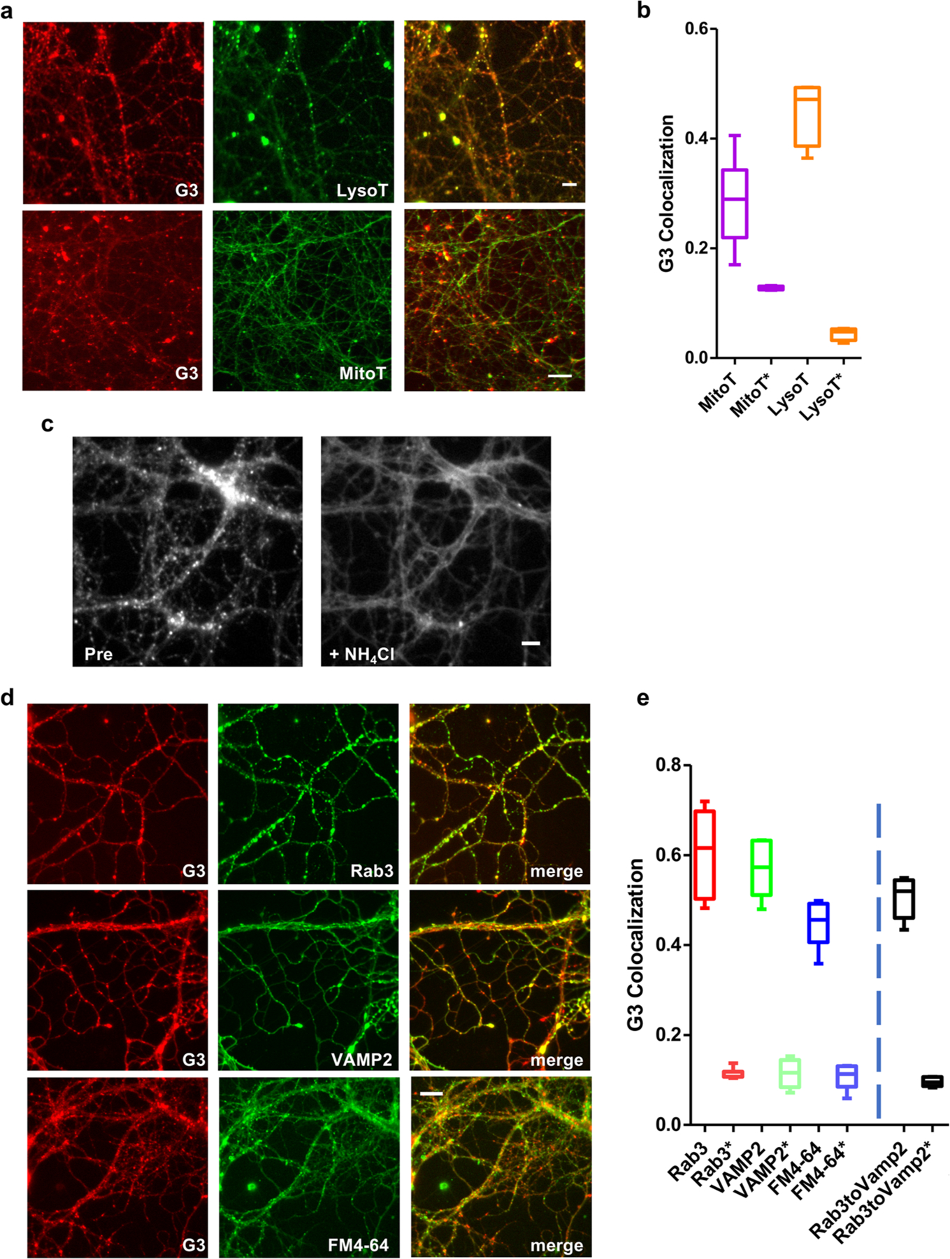
CX-G3 is a pH-sensitive dye that labels synaptic vesicles in cultured neurons. (a) Images showing colocalization of CX-G3 with LysoTracker Red and MitoTracker Red in hippocampal neurons. Size bar is 10 μm. Note that MitoTracker images were acquired using a different imaging system (see Methods). (b) Graph of CX-G3 colocalization with LysoTracker and MitoTracker, defined as fraction of CX-G3 puncta which contain fluorescent marker. Colocalization of G3 with these dyes in randomized images (depicted with *) are included for comparison (n = 2–4 images per batch, acquired from two independent batches of neurons). (c) Images of CX-G3-labeled neurons before (pre) and after deacidification with 50 mM NH4CL (d) Images showing colocalization of CX-G3 with mCh-tagged SV markers (Rab3 or VAMP2) or FM4–64 to label active terminals. Size bar is 10 μm. (e) Graph of CX-G3 colocalization with SV markers in cultured hippocampal neurons, defined as fraction of CX-G3 puncta which contain fluorescent marker. Colocalization of G3 with Rab3, VAMP2, and FM in randomized images (depicted with *), and colocalization of mCh-Rab3 with endogenous VAMP2 via immunostaining are included for comparison (n = 2–4 images per batch, acquired from two independent batches of neurons).
