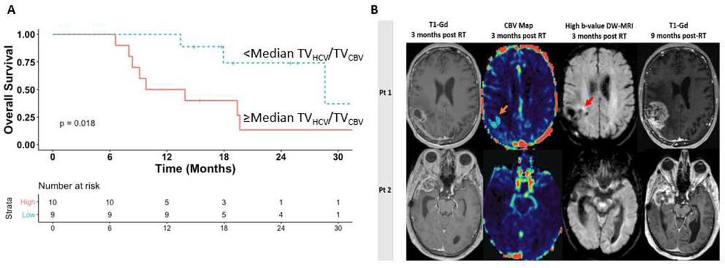
Figure 2.
Panel A. Kaplan-Meier curves for overall survival demonstrating significantly improved outcomes among patients with <median residual hypercellular/hyperperfused tumor volumes versus ≥median 3 months post-chemoradiation. Panel B. At 3 months post-radiation, standard contrast-enhanced MRI in 2 different patients (left column) was interpreted as stable disease and the patients were continued on maintenance temozolomide. Persistent hyperperfused tumor (TVCBV orange arrow, 2nd column) and hypercellular tumor (TVHCV red arrow, 3rd column) is detectable medially using multiparametric MRI at this time point in patient 1. In contrast, patient 2 did not have persistent residual hyperperfused/hypercellular disease. Approximately six months later, both patients developed asymptomatic worsening of their contrast-enhanced MRI prompting resection. Patient 1 had pathologically confirmed tumor progression. Patient 2 had gliosis/inflammation without residual tumor. Gd=Gadolinium; CBV=Cerebral blood volume; DW-MRI=Diffusion-weighted magnetic resonance imaging; RT=Radiation therapy

