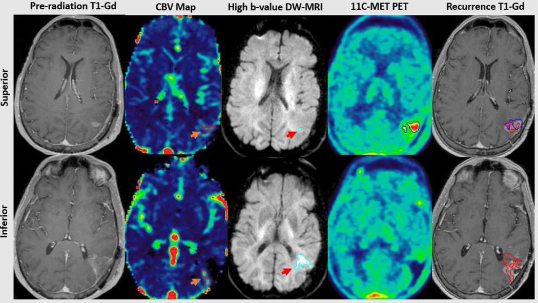
Figure 3.
Following apparent gross total resection (contrast-enhanced MRI, far left), residual tumor is demonstrated adjacent to the superior and inferior portions of the cavity. The superior residual consists of hyperperfused (orange arrow, top 2nd panel) and a small volume of hypercellular tumor (red arrow, top 3rd panel), concordant with a region of metabolic abnormality detected using 11C-Methionine PET (top 4th panel). The inferior residual consists of predominantly hypercellular (red arrow, bottom 3rd panel) and some hyperperfused tumor (orange arrow, bottom 2nd panel), without associated metabolic abnormality (bottom 4th panel). Pathologically confirmed central tumor recurrence was concordant with all 3 advanced imaging tumor volumes superiorly (red and blue, upper far right panel) and the hypercellular/hyperperfused tumor inferiorly (red, bottom far right panel). Gd=Gadolinium; CBV=Cerebral blood volume; MET-PET=Methionine Positron Emission Tomography

