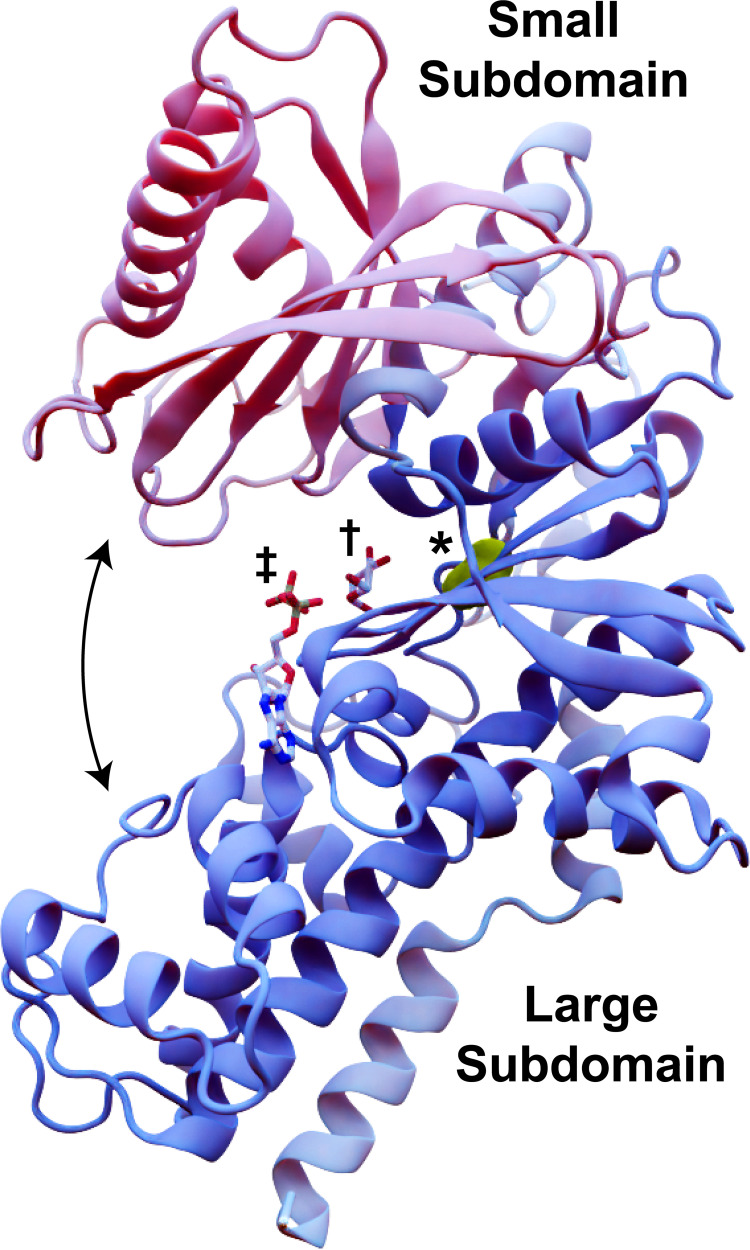
Fig 1. ScHxk2 structure and global dynamics.
The mostly helical large subdomain and the ɑ/β small subdomain are shown in blue and pink, respectively (PDB 1IG8). The curved arrows indicate the approximate motion of domain closure. The location of residue 238 is shown in yellow and marked with an asterisk. To indicate the location of the glucose- and ATP-binding pockets, we superimposed crystallographic glucose (dagger) and ADP (double dagger) molecules from structures of HsHk1 (human, PDB 4FPB) and OsHxk6 (rice, PDB 6JJ8), respectively.

