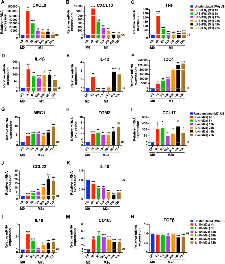
Fig 1. Time depended changes in the expression of macrophage polarization markers at mRNA level.
Primary human MDMs (M0) were left unstimulated or were polarized to M1 (100 ng/mL LPS, 20 ng/mL IFNγ), M2a (20 ng/mL IL-4), or M2c (20 ng/mL IL-10) for 4, 8, 12, 24, 48, or 72 h. The expression of (A-F) CXCL9, CXCL10, TNF, IL-1β, IL-12, and IDO1 mRNA for M1 polarization; of (G-K) MRC1, TGM2, CCL17, CCL22 and IL-10 mRNA for M2a polarization; and of (L-N) IL-10, CD163 and TGFβ mRNA for M2c polarization were analyzed by qPCR. Data shown are mean ± SEM of biological replicates of 3 independent donors. Polarized macrophages (M1, M2a, or M2c) at all time points were compared with unstimulated (US) M0 macrophages. Trends for individual donors are depicted in S1 Fig. Statistical analyses were performed with repeated measures ANOVA, * p < 0.05; ** p < 0.01; *** p < 0.001 (Statistical comparison of all time points are shown in the S1 Table).

