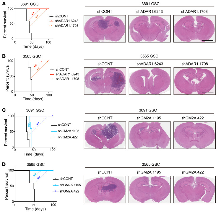
Figure 8. Targeting ADAR1 or GM2A attenuates in vivo tumor growth.
(A) Survival analysis of NSG mice bearing intracranially implanted patient-derived 3691 GSCs transduced with shCONT or 1 of 2 non-overlapping shADAR1s (left panel). Statistical significance was determined by log-rank (Mantel-Cox) test. GSCs were implanted intracranially into NSG mice, and tumor formation was determined by H&E staining (right panel). Scale bar: 2 mm. n = 5 per group, 2 biological replicates. (B) Survival analysis of NSG mice bearing intracranially implanted patient-derived 3565 GSCs transduced with shCONT or 1 of 2 non-overlapping shADAR1s (left panel). Statistical significance was determined by log-rank (Mantel-Cox) test. GSCs were implanted intracranially into NSG mice, and tumor formation was determined by H&E staining (right panel). Scale bar: 2 mm. n = 5 per group, 2 biological replicates. (C) Survival analysis of NSG mice bearing intracranially implanted patient-derived 3691 GSCs transduced with shCONT or 1 of 2 non-overlapping shGM2As (left panel). Statistical significance was determined by log-rank (Mantel-Cox) test. GSCs were implanted intracranially into NSG mice, and tumor formation was determined by H&E staining (right panel). Scale bar: 2 mm. n = 5 per group. (D) Survival analysis of NSG mice bearing intracranially implanted patient-derived 3565 GSCs transduced with shCONT or 1 of 2 non-overlapping shGM2As (left panel). Statistical significance was determined by log-rank (Mantel-Cox) test. GSCs were implanted intracranially into NSG mice, and tumor formation was determined by H&E staining (right panel). Scale bar: 2 mm. n = 5 per group. **P < 0.01.

