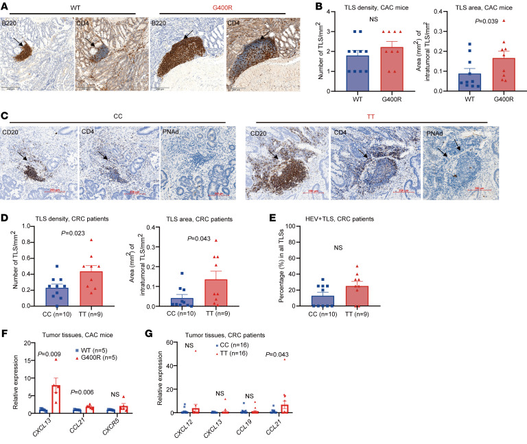
Figure 5. hIgG1-G396R promotes the formation of tertiary lymphoid structures in tumor tissues.
(A) Microphotographs of representative tertiary lymphoid structures (TLSs) located in the tumor section of CAC-induced mice. Black arrows indicate TLSs. Scale bars: 200 μm. (B) IHC showing the numbers and relative areas of TLSs in CAC-induced mice. (C) Representative IHC of TLSs and PNAd+ TLSs within the tumor specimens from CRC patients, as indicated by black arrows. Scale bars: 200 μm. (D and E) The numbers of TLSs per mm2, relative TLS area, and the percentages of HEV+ TLSs for each genotype are shown. (F) The expression levels of several chemokine genes associated with TLS formation in the colon tumor tissues from CAC-induced mice by RT-qPCR assays. (G) The expression levels of several chemokine genes in the tumor specimens from CRC patients quantified by RT-qPCR assays. One of 3 representative experiments is shown (F and G). Statistical significance was determined using an unpaired, 2-tailed Student’s t test. Data are presented as the mean ± SEM.

