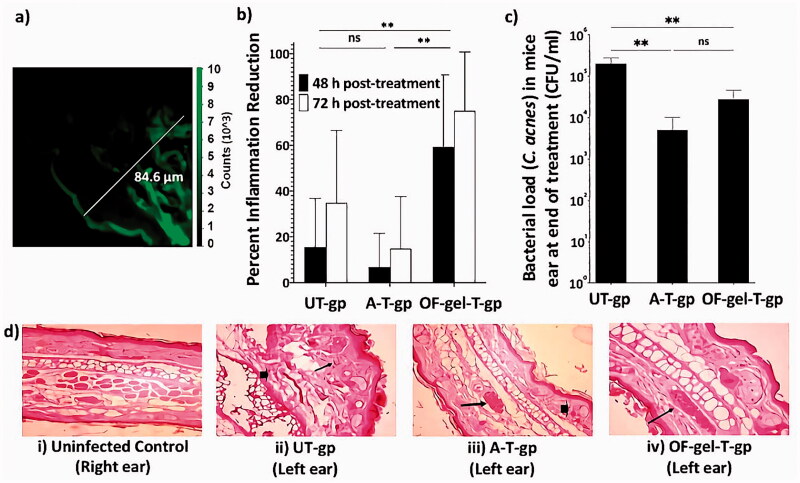
Figure 5.
(a) The flourescence photomicrograph of mouse skin taken using confocal laser scanning microscopy. (b) Percent inflammation reduction after 48 h and 72 h in UT-gp, A-T-gp, and OF-gel-T-gp. (c) Cutibacterium acnes counts in mice ears (represented as colony forming unit (CFU) per mL) at the end of the experiment for UT-gp, A-T-gp, and OF-gel-T-gp. (d) Digital photographs for histopathological evaluation of mice ears at the end of the experiment; (i) healthy control untreated (right ear) where normal histology of ear tissue is shown with absence of inflammatory infiltrate and edema, (ii) infected untreated ear showing necrotic dermatitis characteristics such as sever dermal necrosis (arrow head), malpighian layer atrophy, and lymphocytic infiltrate (arrow), (iii) infected Aknemycin® treated ear showing contact dermatitis characteristics such as dermal hemorrhage (arrow head) and congested blood vessel (arrow), and (iv) infected OF-gel treated ear showing normal histology of mouse ear tissue with basal layer and congested dermal blood vessel (arrow). Ear tissue sections were stained with hematoxylin and eosin stain and all images are 400× magnification. [UT-gp: untreated group, A-T-gp: Aknemycin® treated group and OF-gel-T-gp: Optimized formula-gel treated group]

