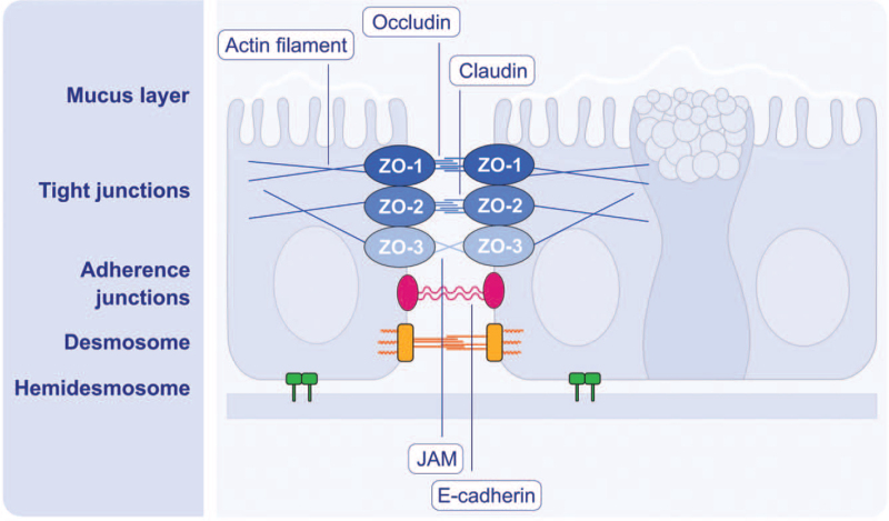
Figure 4.
Schematic diagram of structure and molecular components of airway epithelial barrier. The mucus secreted by goblet cells forms the superficial mucus layer. The junctional structures between epithelial cells from the surface to base are tight junction (TJ), adherence junction (AJ), and desmosomes. TJs, located nearest to the epithelial surface, are key regulators of paracellular permeability depending on the size and ionize of molecules. TJs are constituted of transmembrane proteins including the claudin family (24 claudins), occludin, tricellulin, and junctional adhesion molecules, and the major TJ-associated cytoplasmic proteins are ZO-1, ZO-2, and ZO-3. AJs are located directly below TJs and composed of cadherin-catenin complexes. AJs provide intercellular adhesion to maintain epithelial integrity and perform multiple functions, such as initiation and stabilization of TJs, regulation of the actin cytoskeleton, intracellular signaling, and transcriptional regulation. Desmosomes are located around the midpoint of epithelial cells and contribute to the mechanical stability of airway epithelium due to their strong contact with the intermediate filaments. Hemidesmosomes make the epithelial layer attached to the basal membrane. JAM: Junction adhesion molecule; ZO: Zonula occludens.

