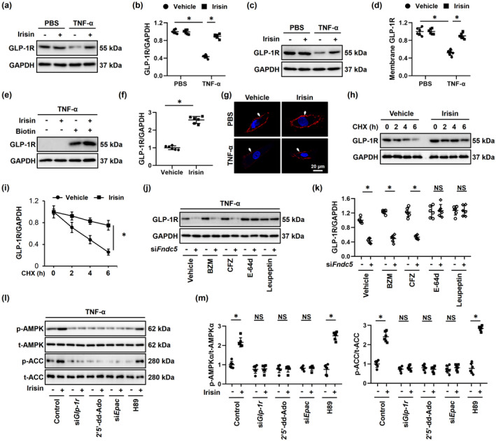
FIGURE 5.

FNDC5 activates AMPKα via blocking the lysosomal degradation of GLP‐1R in vitro. (a‐d) Neonatal rat cardiomyocytes (NRCMs) were treated with 20 nmol/L irisin for 24 h, followed by the stimulation with 100 ng/ml tumor necrosis factor‐α (TNF‐α) or phosphate‐buffered saline (PBS) for an additional 24 h to mimic inflammaging in vitro, and GLP‐1R expressions in total cell lysates or membrane fractions were determined by Western blot (n = 6). (e‐f) NRCMs were subjected to cell surface biotinylation assay, and biotin‐labeled GLP‐1R expression in cell surface was determined by Western blot and normalized to GAPDH in cell lysates. (n = 6). (g) Immunofluorescence staining of GLP‐1R to determine the membrane localization in NRCMs (n = 6). (h‐i) Cells were pretreated with 20 nmol/L irisin for 24 h, followed by the stimulation with 100 ng/ml TNF‐α for an additional 24 h to mimic inflammaging in vitro, and then, cycloheximide (CHX, 10 µmol/L) was used to inhibit protein synthesis at indicated times. GLP‐1R expression in cell lysates was determined by Western blot (n = 6). (j‐k) NRCMs were pretransfected with siFndc5 (50 nmol/L) for 4 h and then maintained for an additional 24 h. To clarify FNDC5‐associated degradation of GLP‐1R, bortezomib (BZM, 0.1 μmol/L), carfilzomib (CFZ, 1 μmol/L), E‐64d (100 nmol/L), or leupeptin (100 µmol/L) was used to inhibit proteasome‐ or lysosome‐mediated degradation. Also, GLP‐1R expression in cell lysates was determined by Western blot. (l‐m) NRCMs were treated with 20 nmol/L irisin for 24 h and then stimulated with 100 ng/ml tumor necrosis factor‐α (TNF‐α) for an additional 24 h. To suppress GLP‐1R, adenylyl cyclase (AC), exchange protein directly activated by cAMP (EPAC), or protein kinase A (PKA), cells were treated with siGlp‐1r (50 nmol/L), 2’,5’‐dideoxyadenosine (2’5’‐dd‐Ado, 200 μmol/L), siEpac (50 nmol/L), or H89 (10 μmol/L). Subsequently, AMPKα and downstream acetyl CoA carboxylase (ACC) phosphorylation in murine hearts were determined (n = 6). Values represent the mean ±standard deviation. *p < 0.05 versus the matched group. NS indicates no significance
