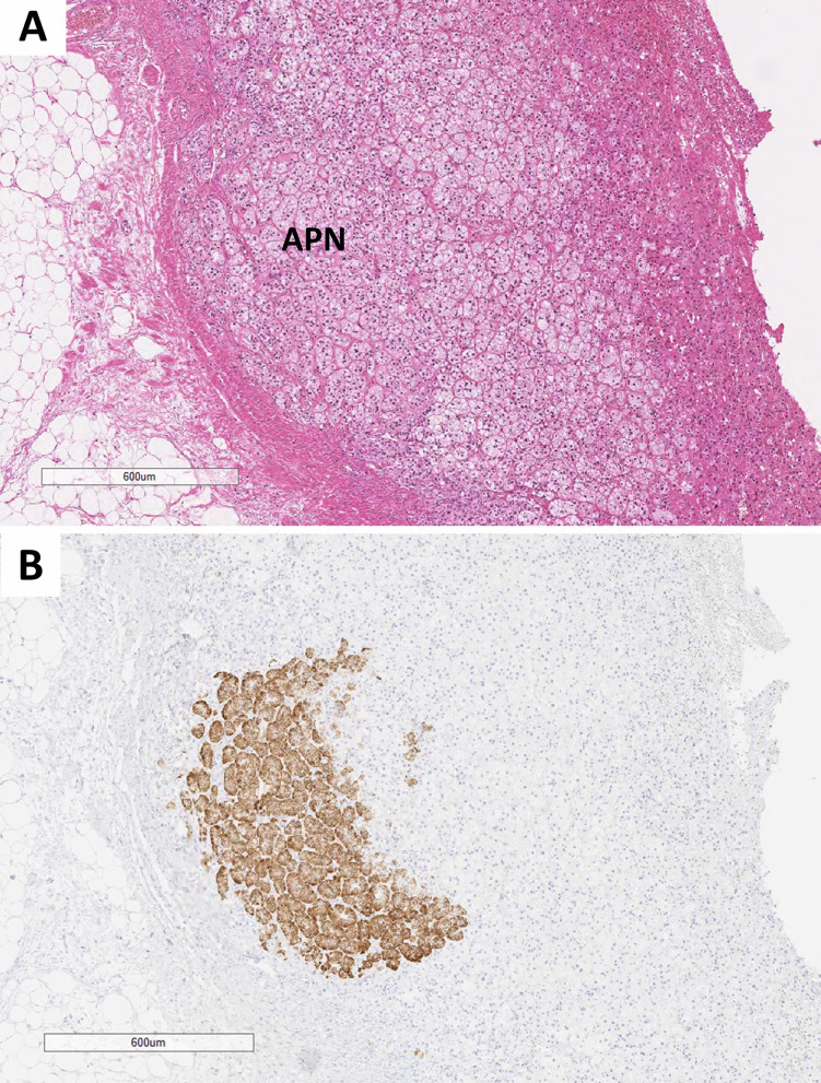
Fig. 14.
Aldosterone-producing nodule. This composite photomicrograph illustrates a morphologically distinct well-delineated adrenal cortical nodule (N) enriched in lipid-rich adrenal cortical cells (A). This nodule measures 1.3 mm. CYP11B2 shows a gradient reactivity with a more intense reactivity at the outer part of the nodule (B)

