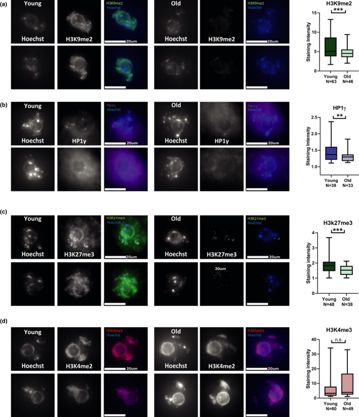
FIGURE 2.

9‐month‐old oocytes lose heterochromatin marks: (a–d) In situ staining images and staining intensity analysis for chromatin marks and proteins in oocytes from young (2‐month‐old) and old (9‐month‐old) females. Oocytes were stained and imaged during prophase I arrest by in situ immunofluorescence (see Methods). Intensity of staining was quantified for each image, and statistical analysis was performed on the numerical values (see Methods). Constitutive heterochromatin marks and proteins were measured by staining for (a) H3K9me2 (t test p < 0.0001, animals used for experiment N young = 4, N old = 4) and (b) HP1γ (t test p = 0.0146, animals used for experiment N young = 3, N old = 3). (c) To examine facultative heterochromatin oocyte were stained and imaged for H3K27me3(t test p = 0.0005, animals used for experiment N young = 3, N old = 5) (d) Euchromatin staining was performed by staining for the marker H3K4me3 (t test p = 0.657, animals used for experiment N young = 3, N old = 6) (e–f)
