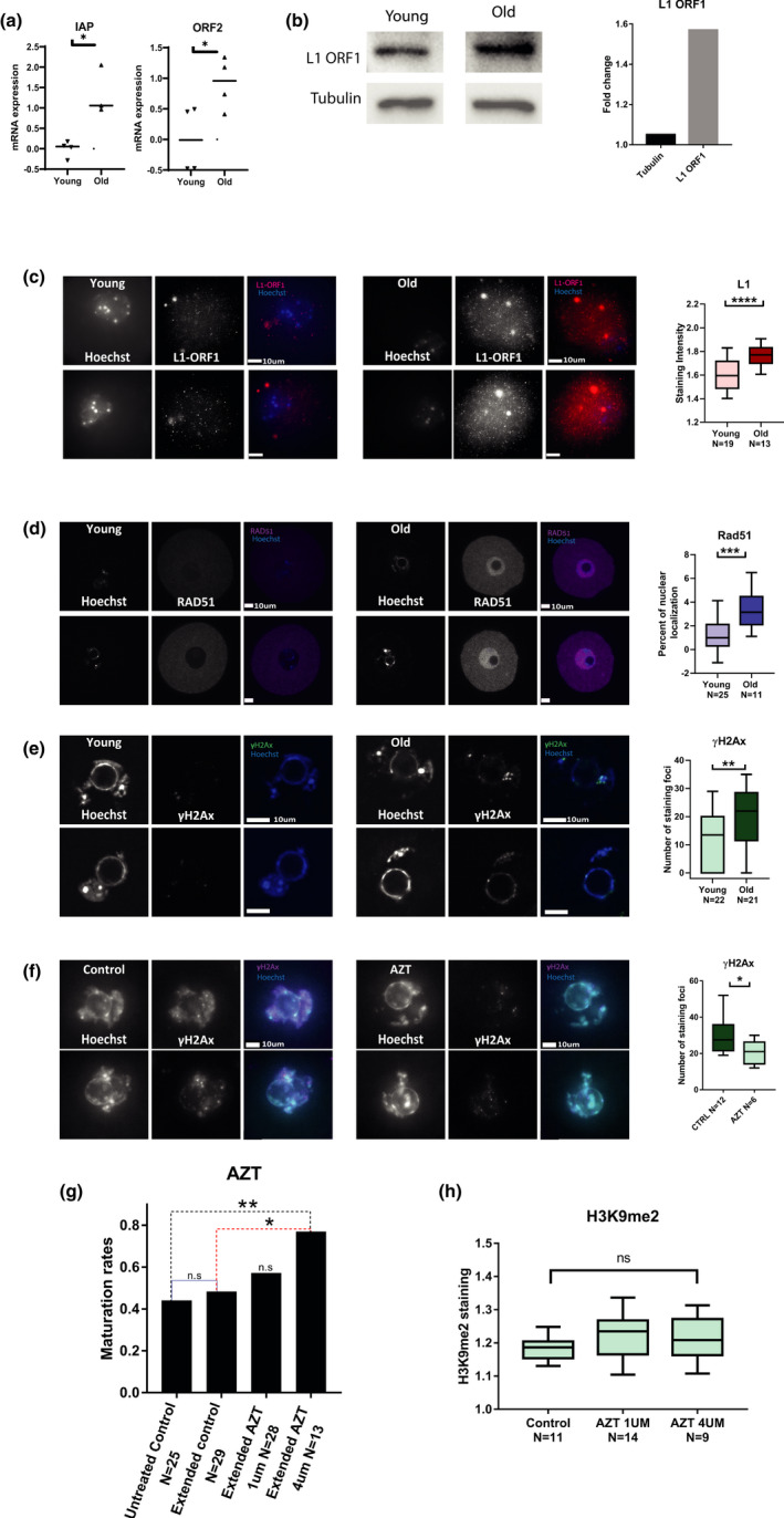
FIGURE 4.

Old oocytes show elevated activity of retrotransposons L1 and IAP and increased DNA damage: (a) Transcriptional retrotransposon activity was measured using qRT‐PCR for L1 retrotransposon (MW p = 0.01, animals used for experiment N young = 10, N old = 10) and IAP elements mRNA (MW p = 0.03, animals used for experiment N young = 10, N old = 10) (see Methods) in young (2‐month‐old) and old (9‐month‐old) prophase I‐arrested mouse oocytes. (b) Upregulation of L1 retrotransposon protein was measured by Western blotting for L1‐ORF1p in from young (2‐month‐old) and old (9‐month‐old) prophase I‐arrested mouse oocytes. (N young = 6, N old = 8) (c) Upregulation of L1 retrotransposon activity was measured by in situ staining for L1‐ORF1p in from young (2‐month‐old) and old (9‐month‐old) prophase I‐arrested mouse oocytes. (Shapiro Wilk p < 0.05, t test p < 0.0001, N young = 3, N old = 3). (d) Accumulation of DNA damage with age was assessed by in situ staining for Rad51 and quantification of its nuclear localization (see Methods) in young (2‐month‐old) and old (9–month‐old) prophase I‐arrested mouse oocytes (MW p < 0.0005, N young = 3, N old = 5). (e) Another measure for DNA damage was performed by in situ staining for γH2AX (MW p = 0.0085, N young = 3, N old = 5) that was quantified by the number of nuclear foci in old and young prophase I‐arrested mouse oocytes (see Methods). (f) To investigate the phenotype of aged oocytes after retroviral activity inhibition, oocytes were treated with AZT and then stained for γH2A and the number of nuclear foci in AZT treated and untreated control old prophase I‐arrested oocytes was quantified (MW p < 0.0274 animals used for experiment N = 3) (g) The maturation efficiency of old oocytes treated with two different doses of AZT and matured in vitro was measured by proportion of oocytes which successfully completed MII after treatment (Z test p = 0.0102) (see Methods). Because incubation with the drug required longer incubation in IBMX than normal, an extended incubation without AZT was also added as control (h) Quantification of in situ staining intensity for H3K9me2 in control old oocytes and old oocytes treated with two different doses of AZT (see Methods)
