FIGURE 6.

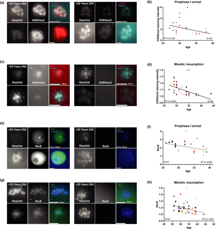
Human oocytes show a reduction of the H3K9me2 heterochromatin marker, and REC8 meiotic cohesion marker with age: To study the changes in heterochromatin with age in Human oocytes, human GV‐arrested oocytes were stained by immunofluorescence for H3K9me2 (see text and Methods). Oocytes were retrieved from 33 women, with average of 2.2 oocyte per woman. Analysis was performed separately for oocytes that were stained during prophase I arrest or arrested later during meiosis. (a) Examples of stained (for H3K9me2) prophase I‐arrested oocytes from patients under 35‐year‐old and over 35‐year‐old (b) Oocytes arrested during meiosis also show a reduction of signal intensity with age with a linear correlation, (N = 28, F test p = 0.0008). The oocytes of each patient are in a different color (for a given age, several patients with more than one oocyte may be represented). (c) Examples of stained (for H3K9me2) prophase I oocytes after meiotic resumption, from patients under 35‐year‐old and over 35‐year‐old. (d) Prophase I oocytes show a reduction of signal intensity with age with a linear correlation. (N = 21, F test p = 0.03) (e) Examples of in situ staining of prophase I‐arrested human oocytes (N = 21) for Rec8 meiotic cohesion subunit. (f) These oocytes show a linear reduction of signal intensity with age (F test p = 0.02). (g) Examples of in situ staining of human prophase I oocytes after meiotic resumption (N = 47) for Rec8. (h) These oocytes show a linear reduction of signal intensity with age (F test p = 0.01)
