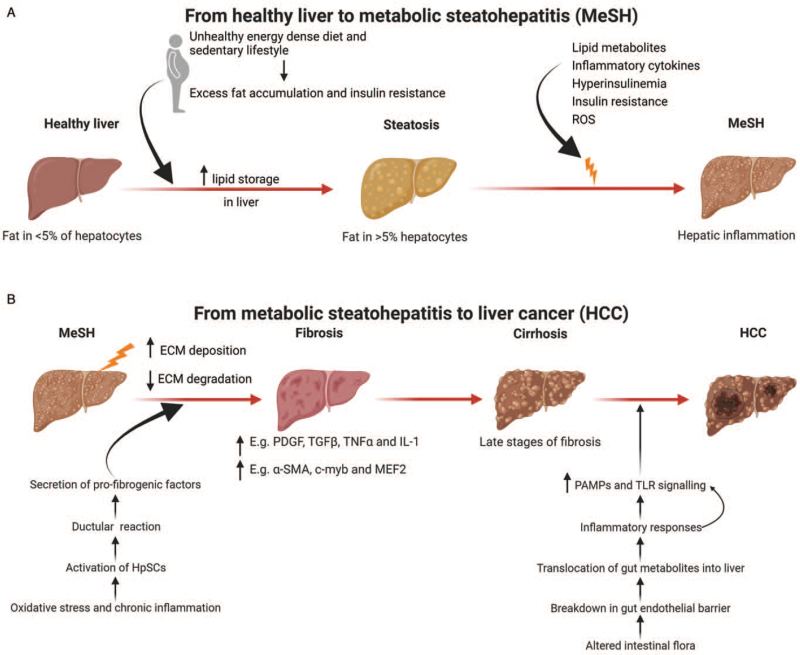
Figure 1.
Disease progression from a healthy liver to HCC. (A) A sedentary lifestyle, unhealthy, energy dense diets, and reduced physical activity are major contributors to obesity. This leads to insulin resistance and excess liver fat accumulation. Excess lipid storage in the liver in predisposed individuals triggers hepatic inflammation or MeSH. (B) Persistent MeSH leads to an increase in ECM deposition and a decrease in matrix degradation resulting in liver fibrosis. Hepatic fibrosis is driven by a variety of inflammatory molecules. Cirrhosis is late-stage liver fibrosis and is the substrate in most cases for the development of liver cancer, though in MAFLD, HCC can develop in the absence of cirrhosis. Adapted from “Non-Alcoholic Fatty Liver Disease (NAFLD) Spectrum”, by BioRender.com (2021). Diagram retrieved from https://app.biorender.com/biorender-templates. α-SMA: Alpha-smooth muscle actin; ECM: Extracellular matrix; HpSCs: Hepatic stem/progenitor cells; HCC: Hepatocellular carcinoma; IL-1: Interleukin-1; MAFLD: Metabolic (dysfunction) Associated Fatty Liver Disease; MEF2: Myocyte enhancer factor 2; MeSH: Metabolic steatohepatitis; NAFLD: Non-alcoholic fatty liver disease; PDGF: Platelet-derived growth factor; ROS: Reactive oxygen species; TLR: Toll-like receptor; TGFβ: Transforming growth factor beta; TNFα, tumor necrosis factor alpha.

