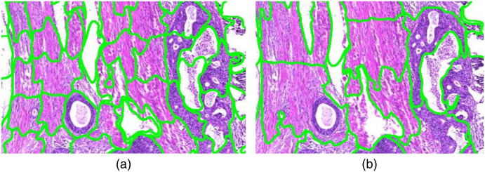
Fig. 5.
(a) Superpixel segmentation result with size of 3.600 pixels compared to (b) the segmentation result with a size of 10.000 pixels. The segmentation results on the left side fits better to the tumor cell boundaries but in both images necrotic areas within the tumor are not always detected as separate regions.

