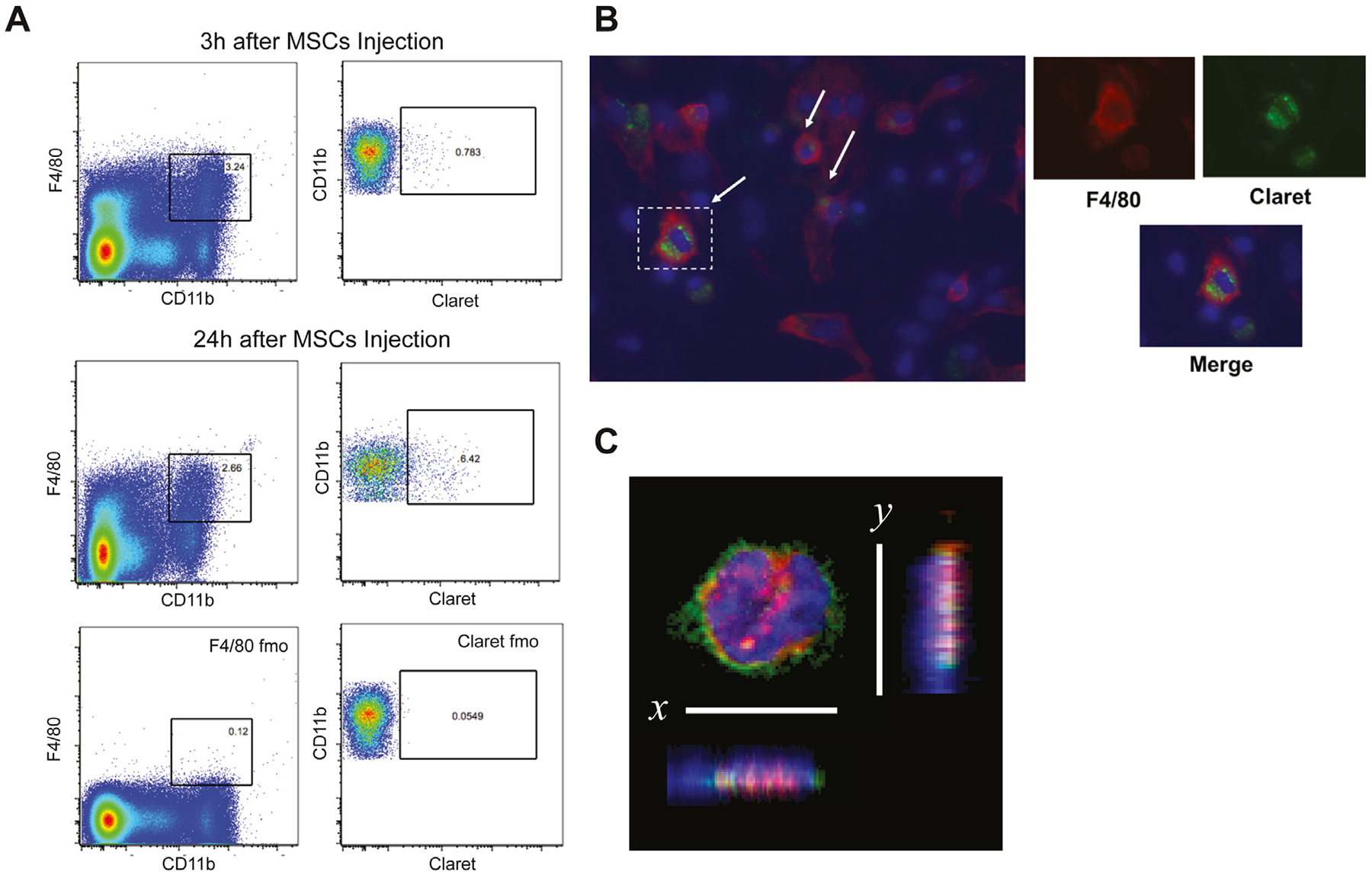
Figure 7.

Analysis of spleen macrophages after MSC injection. (A) After intravenous injection of Claret-labeled MSCs, spleen cells were isolated and labeled with F4/80 and CD11b antibodies. Representative example of flow cytometric analysis at 3 h and 24 h in macrophage spleen cell population. After gating on macrophages (F4/80+CD11b+), Claret+ cells were identified according to the expression of Claret on this macrophage population. (B) Following isolation of spleen cells, cells were cultured for 3 days and stained with F4/80 antibody. Claret staining was found inside the macrophages. Smaller panels represent enlarged views of dotted rectangles. DAPI (blue) stained nucleus and anti-F4/80 antibody (red) stained macrophages. (C) MSCs were labeled with DiI membrane stain (red) and administered intravenously 4 h before animals were killed. Spleen cell suspension was stained with F4/80 antibody (green) and DAPI (blue), cytospinned and imaged with confocal microscopy. The image was digitally rotated along the x- or y-axis to demonstrate that the DiI signal (engulfed MSCs) was within the macrophage (magnification ×63). DAPI, 4’,6-diamidino-2-phenylindole; fmo, fluorescence minus one.
