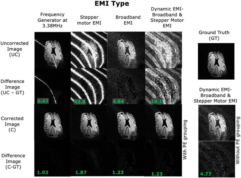
Figure 6: EDITER-corrected 2D images of a brain slice phantom acquired in the portable 80mT scanner.
Results are shown for the 4 introduced EMI sources describe in Fig. 5. Top to bottom: Uncorrected image, Difference images between uncorrected image and ground truth, EDITER-corrected image using all EMI detectors and Difference images between corrected image and ground truth. The RMSE of the difference images is shown in the bottom left. For the dynamic EMI source (SM+BB), we show the EDITER correction with dynamic PE grouping and without grouping - where a single impulse response is calculated for all PE lines.

