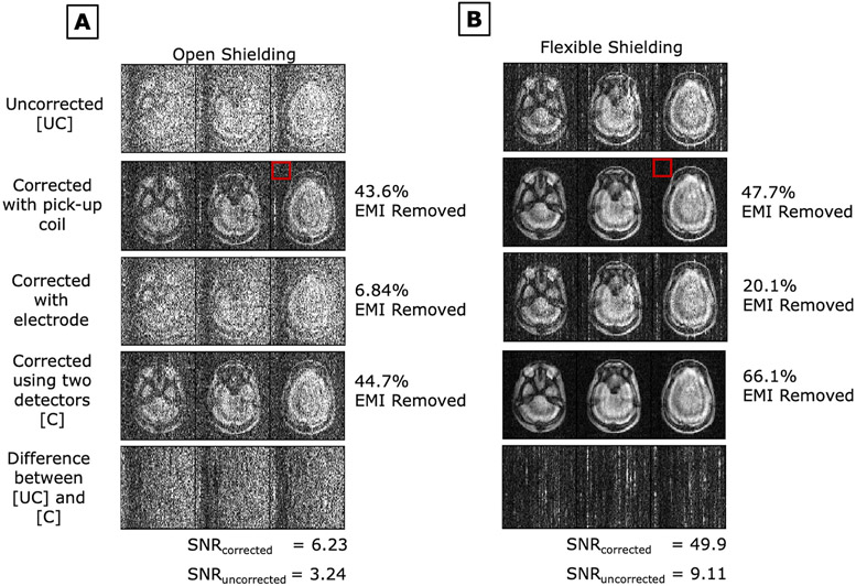
Figure 7:
PD weighted images of an anthropomorphic head phantom in the 47.5mT scanner’s A) open shielding configuration setup and B) flexible shielding configuration setup (with draped copper mesh). EDITER correction is shown using each EMI detector individually and together. Percent EMI removed by the EDITER method using Eq.4 is indicated on the right. The red box indicates the region outside the object that was used for this measurement. SNR for both the uncorrected and corrected images are shown.

