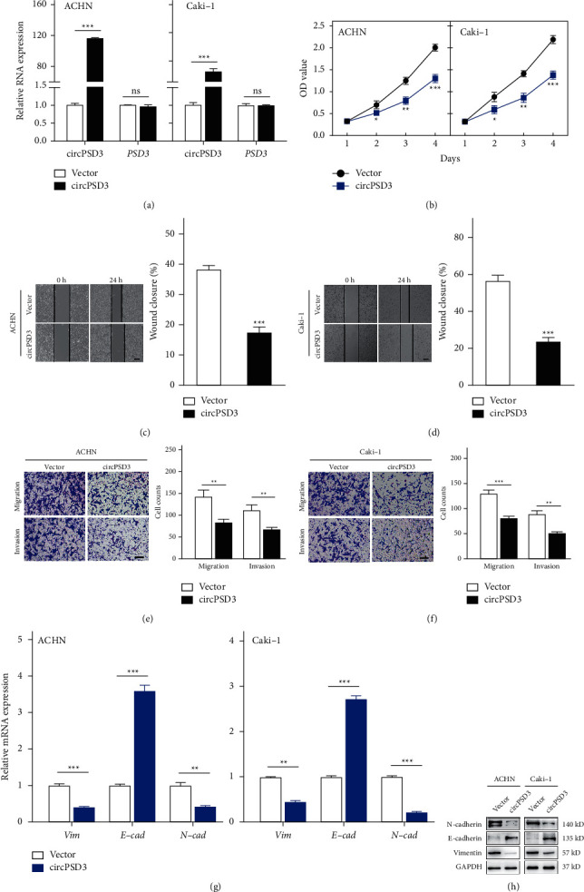
Figure 3.

Overexpression of circPSD3 inhibits the migration and invasion of ccRCC cells. (a) qRT-PCR analysis of circPSD3 and PSD3 RNA expressions in ACHN and Caki-1 cells infected with a circPSD3 overexpression of lentivirus or vector control lentivirus. (b) Cell Counting Kit-8 assay of the effect of circPSD3 on the survival of the indicated ccRCC cells with stable circPSD3 overexpression. (c, d) Wound healing assays were performed to analyze the migration capacity of the indicated ccRCC cells. The dashed lines indicate the edge of migrating cells. Scale bars, 200 μm. (e, f) Transwell assays were performed using Matrigel-coated or uncoated inserts to determine the migration and invasion capacities of the indicated ccRCC cells. Scale bar, 100 μm. (g, h) qRT-PCR and Western blot analyses of the mRNA and protein levels of E-cadherin, N-cadherin, and vimentin in the indicated ccRCC cells. GAPDH was used as a loading control. The data are presented as mean ± SD based on triplicate independent experiments. ∗P < 0.05; ∗ ∗P < 0.01; and ∗ ∗ ∗P < 0.001; and NS, no significance.
