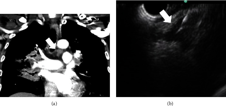
Figure 2.

A 53-year-old male patient. (a) Left lower paratracheal lymphadenopathy (arrow) on CT scan. (b) On EUS view, triangle-shaped hypoechoic lymph node (arrow) was observed in station 4L. The EUS-guided biopsy was performed. CT: computed tomography; EUS: endoscopic ultrasound.
