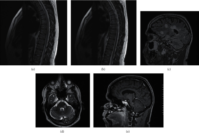
Figure 3.

((a), (b)) There are at least 2 definite high signal intensity plaques on the sagittal T2W1 seen at D6-7, D9-10 levels (arrow). ((c), (d)) Numerous bilateral variable-sized, different-shaped plaques of high signal intensity on T2W1/FLAIR seen mainly in the deep white matter at periventricular location and centrum semiovale, perpendicular callososeptal lesions, cerebellar peduncles, and cerebellar white matter. (e) Incomplete ring enhancement lesions seen in the left deep periventricular frontal lobe and left cerebellar peduncle.
