Figure 1.
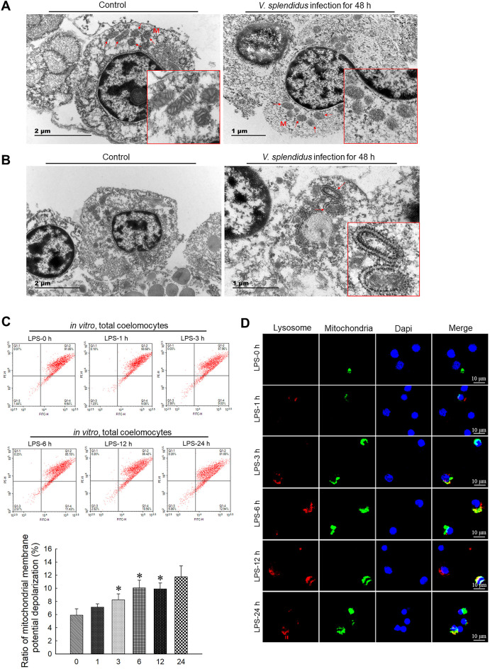
Vibrio splendidus infection and LPS challenge induced mitochondrial damage and mitophagy in coelomocytes
A: Representative TEM images of morphology of damaged mitochondria (arrowhead) in coelomocytes of A. japonicus infected by V. splendidus for 48 h. B: Representative TEM images of autophagosomes engulfing mitochondria (arrowhead) in coelomocytes of A. japonicus infected by V. splendidus for 48 h. C: ΔΨm of coelomocytes after LPS treatment for 0, 1, 3, 6, 12, and 24 h determined by JC-1 staining and flow cytometry. *: P<0.05;**: P<0.01,n=3. D: Immunofluorescence colocalization analysis of Lyso-Tracker Red with Mito-Tracker Green in coelomocytes after LPS exposure for 0, 1, 3, 6, 12, and 24 h, with DAPI (blue) staining to identify nuclei. M: Mitochondria.

