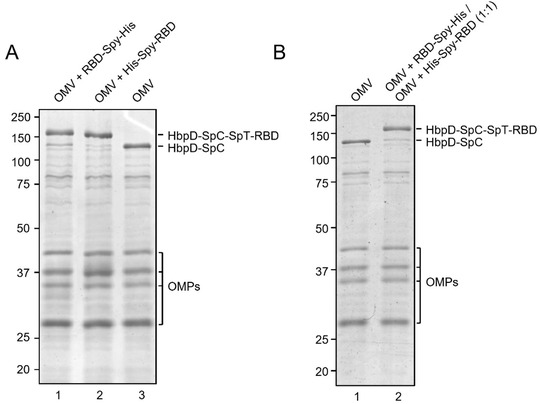
FIGURE 2.

(A) Assessment of efficiency of SpyTag/SpyCatcher coupling of RBD onto HbpD of OMVs. RBD‐Spy‐His and His‐Spy‐RBD were coupled to Hbp‐SpyCatcher OMVs. Proteins of conjugated and non‐conjugated OMVs were separated by SDS‐PAGE and stained with Coomassie Brilliant Blue. RBD‐HbpD appears as a ∼160 kDa band, while free HbpD is seen as a ∼125 kDa band. Densitometry suggested that approximately 90% or more of HbpD was coupled with RBD in the conjugated populations compared with unconjugated OMVs (rightmost lane). Other outer membrane proteins of OMVs (OMPs) are indicated; (B) Coomassie Brilliant Blue staining of SDS‐PAGE gel containing non‐conjugated OMVs and a 1:1 mixture of RBD‐Spy‐His and His‐Spy‐RBD‐coupled OMVs
