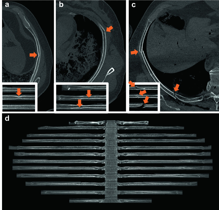
Fig. 1.
Rib unfolding. (a) Multi-planar view of a so-called buckle rib fracture (subgroup of incomplete rib fractures). Inset lower left: Rib unfolding view of a buckle rib fracture with no interrupted cortical line but obvious kinking. (b) Multi-planar view of an incomplete rib fracture; outer cortical line interrupted. Inset lower left: Rib unfolding view of an incomplete rib fracture with interrupted cortical line. (c) Multi-planar view of two complete rib fractures in the middle and the back of the rib. Inset lower left: Rib unfolding view of three complete rib fractures. In contrast to the two other fractures (a and b), both cortical lines are interrupted in complete fractures. (d) Rib unfolding with no rib fractures and 12 ribs on either side of the vertebral column. The cross-sectional images (a, b, and c) were extracted with bone window settings (center 450 HU, width 1,500 HU), while the window settings used for the unfolded rib images (d) and the insets (a, b, and c) were 1,000 HU for the center and 2,500 HU for the width

