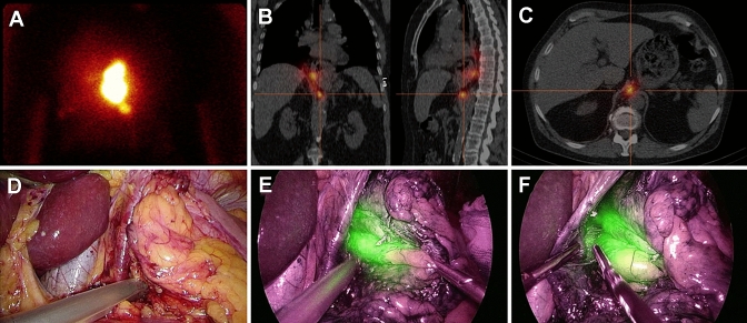
Fig. 1.
Identification of a retrocrural located sentinel node. A: Lymphoscintigraphy 120 min after injection of the hybrid tracer showed the injection site and a sentinel node located below. B + C: This was combined with a SPECT/CT of the thorax and abdomen to detect the exact sentinel node location. D: High radioactivity uptake was confirmed with the laparoscopic gammaprobe during the abdominal phase of surgery. E: The sentinel node was also clearly visualized as indocyanine green positive after switching the camera view to near-infrared. F: Laparoscopic resection of the sentinel node was started while visualized with the near-infrared camera

