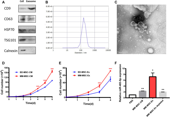
FIGURE 3.
Exosome characterization and function. (A) Exosome-related positive-markers like CD9, CD63, HSP70, TSG101 and negative-markers like Calnexin were detected by Western blot (B) The particle size was determined by Nanosight particle analysis. (C) TEM analysis showed the cup-shaped morphology of exosomes. (D, E) Both MM-MSC conditioned medium (D) and exosomes (E) showed significant pro-proliferative roles in MM cells compared with BD-MSCs. (F) miR-483-5p expression after treatment with conditioned medium or exosomes was tested by qRT-PCR.

