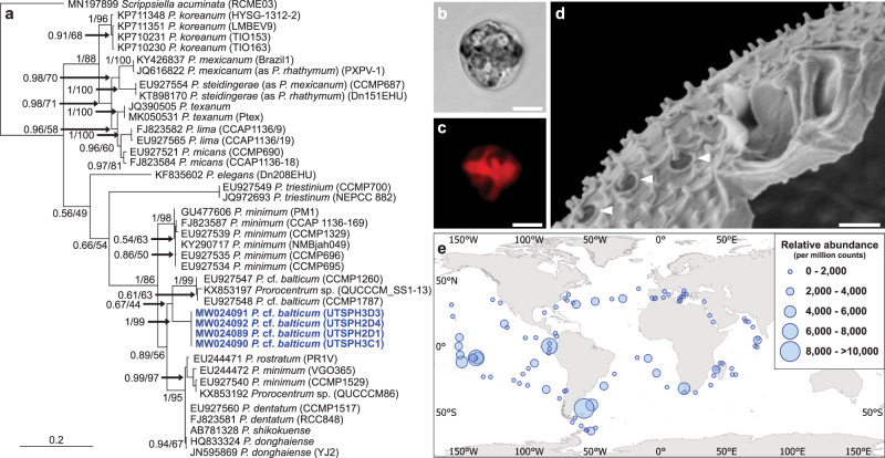
Fig. 1. Identification and distribution of Prorocentrum cf. balticum.
a Maximum Likelihood phylogenetic tree showing alignment of the internal transcribed spacer (ITS) gene region indicating a clear differentiation of the P. cf. balticum strains (blue) from described Prorocentrum species, and the close relation to undefined strains. Accession numbers, species designation, and strain codes are provided for each sequence and values at the nodes represent Bayesian posterior probability and Maximum Likelihood bootstrap support. b, c Light microscope images of P. cf. balticum showing a cell under Differential Interference Contrast (DIC) (b) and peripherally located red-autofluorescent chloroplasts (c). d Scanning Electron Microscope (SEM) image of P. cf. balticum showing distinctive dual wing-like apical projections and unique large pores with emanating large spines (white arrows). Images b, c were taken with an Upright Fluorescence Microscope (Nikon Eclipse Ni, Japan), fitted with a Cy5 620/60 nm ex 700/75 nm em filter and a monochrome camera (Nikon DS-Qi2) under 1000× magnification with oil immersion; scale bar = 5 µm. Scale bar in d = 0.5 µm. e Global biogeography of P. cf. balticum produced using the relative abundance of 18S rDNA sequences from the Tara Oceans amplicon dataset.

