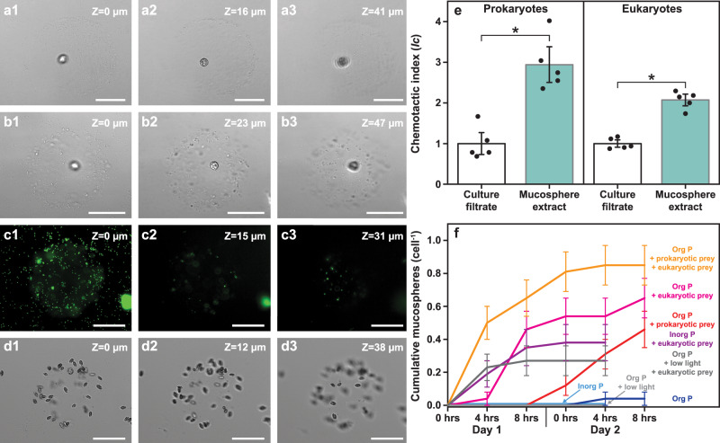
Fig. 3. Detail of mucospheres constructed by Prorocentrum cf. balticum cells to aid prey capture.
a–d Three-dimensional structure of the mucospheres with a Z-stack sequence from the (1) bottom, (2) middle and (3) top, with a mucosphere constructed in the absence of prey (axenic culture) (a), a mucosphere with captured prokaryotes (b), the same mucosphere as in b visualised with the fluorescent stain SYBR green (c), and a mucosphere with captured eukaryotic prey (R. salina) (d). e Chemotactic response (as Chemotactic Index (Ic)) of prokaryotic prey (microbiome present in the xenic P. cf. balticum culture) and eukaryotic prey (R. salina) to mucosphere derived chemicals. Mean ± standard error (n = 5 biologically independent samples). Mucosphere-derived chemicals attracted significantly (*) more prokaryotic and eukaryotic prey than culture filtrate controls (two-sided t-test, p = 9.27e−04 for prokaryotic and p = 4.07e−05 for eukaryotic prey). f Cumulative mucosphere production in conditions with different resource availability observed at 0, 4, and 8 h after illumination (14:10 light:dark cycle) over two days. The calculated values include when a single P. cf. balticum cell produced multiple mucospheres. Mean ± standard error (n = 26 cells; Kruskal–Wallis test, p = 4.06e−12). Images a–d were taken with an Inverted Fluorescence Microscope (Nikon Eclipse Ti), fitted with a Nikon FITC 480/30 nm ex 535/45 nm em filter and a monochrome camera (Nikon DS-QiMc) under 200× magnification. Scale bars = 50 µm.

