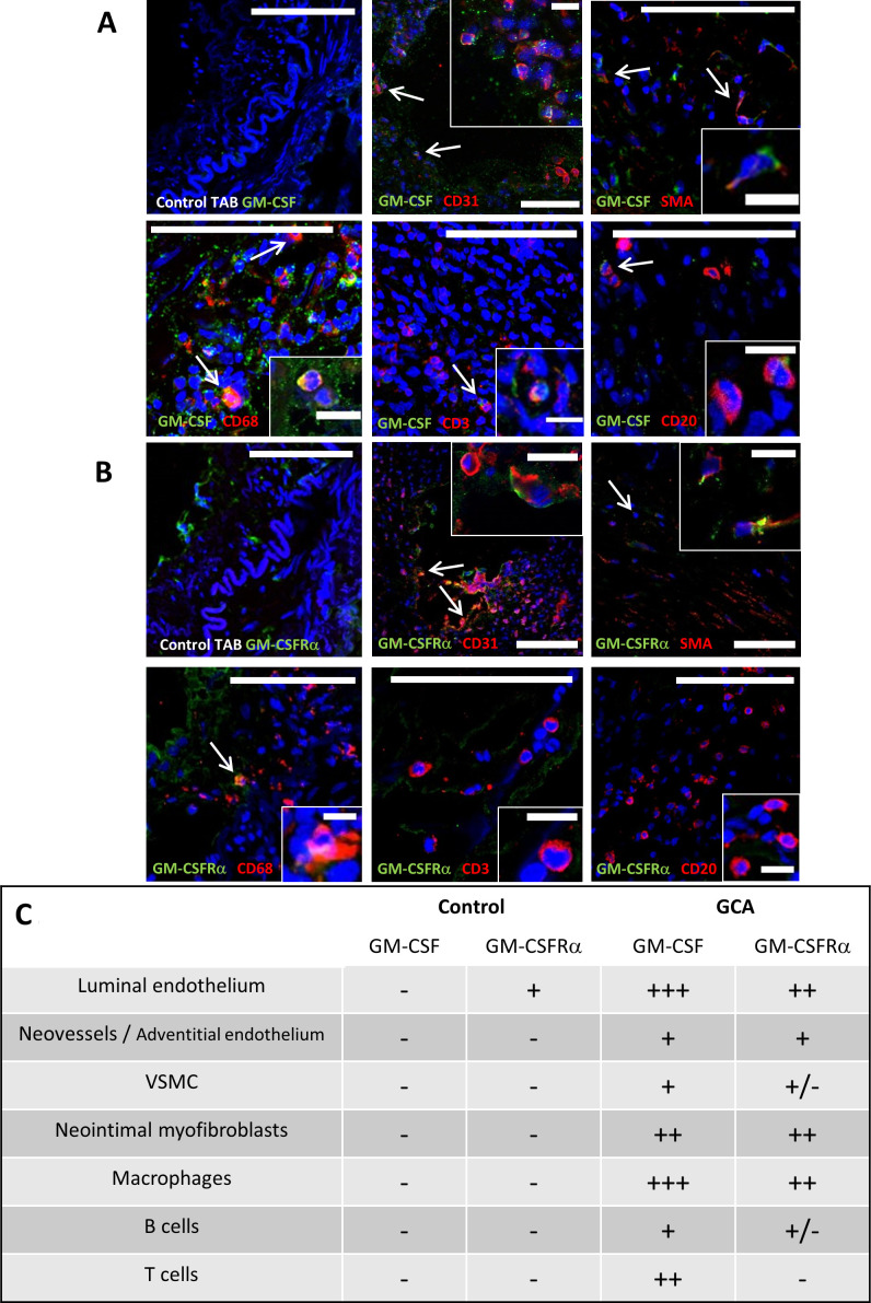
Figure 2.
GM-CSF and GM-CSFRα expression by immune and resident cells. Merged double immunofluorescence staining with anti-GM-CSF (A) or anti-GM-CSFRα (B) antibodies (both in green) and cell surface markers CD68 (macrophages), CD31 (endothelial cells), CD3 (T lymphocytes), CD20 (B lymphocytes) and SMA (identifying vascular smooth muscle cells and myofibroblasts) (all in red) of fresh-frozen temporal arteries from patients with GCA or controls (first panel). Nuclei are stained with DAPI (blue). Co-expression (orange/yellow) is pointed with arrows and insets show magnified double-positive cells (scale bars in figures measure 100 μm and 15 μm for insets). (C) Summary panel of GM-CSF and GM-CSFRα expression by different cell types in three GCA-involved temporal arteries detected by immunofluorescence as in A and B. +++: 50%–100% positive cells; ++: 20%–40% positive cells; +: less than 20% positive cells; +/−: scattered cells; −: negative. DAPI, 4′,6-diamidino-2-phenylindole; GCA, giant cell arteritis; GM-CSF, granulocyte-macrophage colony stimulating factor; GM-CSFRα, GM-CSF receptor alpha chain; SMA, smooth muscle actin; TAB, temporal artery biopsy; VSMC, vascular smooth muscle cells.

