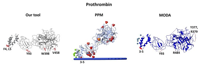
Figure 4.
Comparison of the predictions provided by our model, PPM and MODA for the open form of the prothrombin protein. Our model and MODA suggest an orientation parallel to a putative membrane plane, where Y93 inserts in the membrane which in turn opens the active site of the protease domain (Supplementary Figure S23 available online at http://bib.oxfordjournals.org/). In our model the membrane-penetrating predictions are depicted with red, in PPM the non-protein residues are depicted in CPK representation, and in MODA the membrane-penetrating predictions are depicted with red secondary structure.

