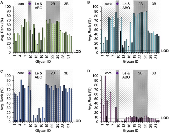
Fig. 4.

Relative average binding intensities comparing the core fucosylated structures (colored bars) with the corresponding noncore fucosylated glycan (black bar) for (A) AOL (green), (B) AAL (cyan), (C) LCA (blue) and (D) PhoSL (fuchsia). All the structures (numbered in the horizontal axis) were also categorized according to common considerations found in the specialized literature: core means short paucimannosidic oligosaccharides; purple diamond refers to distal sialylated glycans; Le and ABO are glycan structures having the Lewis or blood group epitopes in at least one of their antennas; 2B and 3B are di- and tri-antennary glycans, respectively. Complete structures can be seen in Table SIV. Dotted line refers to the LOD calculated for each lectin.
