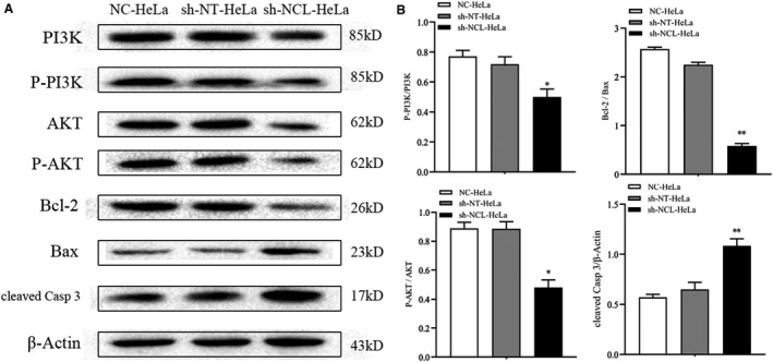
FIGURE 5.

Protein levels of PI3K, AKT, Bcl‐2, Bax, and cleaved‐Caspase 3 were detected by Western blotting. (A) Representative Western blot brands. (B) Quantitative analyses of phospho‐PI3K/PI3K, phospho‐AKT/AKT, Bcl‐2/Bax, cleaved‐Caspase 3. Mean ± SD from three individual experiments. *p < 0.05, **p < 0.01, versus the sh‐NT‐HeLa group
