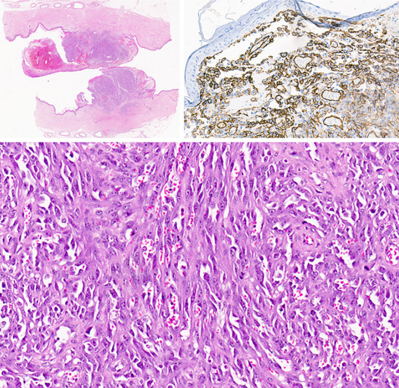
Fig. 3.
Pathology description. Macroscopic examination of the breast showed a polypoid lesion on the skin with ulceration. The low magnification image shows the lesion involving the dermis of the skin and no extension or multifocal foci within the deeper tissues. The tumor morphology shows a vasoformative spindle cell proliferation with red blood cells within the vascular spaces formed by the spindle cells. The tumor cells are positive with CD34 and CD31. In situ hybridization for the MYC gene showed amplification of MYC with a mean copy number of 13.7 copies per nucleus (CEP8 aqua; MYC red). Together the features are indicative of post-radiation angiosarcoma of the breast.

