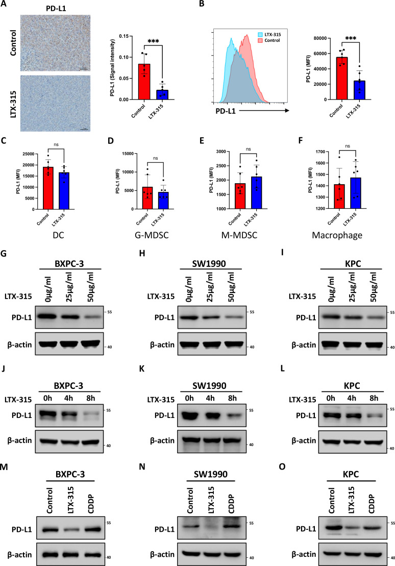
Figure 3.
LTX-315 downregulates PD-L1 expression in pancreatic cancer. (A) Representative images and further quantification of PD-L1 IHC staining in the immunocompetent mice treated with LTX-315. (B–F) Representative images and further quantification of flow cytometry analysis of PD-L1 expression in tumor cells (B), dendritic cells (C), granulocytic-MDSCs (D), monocytic-MDSCs (E), and macrophages (F) in tumors collected from immunocompetent mice. (G–I) LTX-315 inhibition of PD-L1 expression in a dose-dependent manner. PD-L1 expression in BxPC-3 (G), SW1990 (H), and KPC (I) cell treated with LTX-315 at increasing concentrations, individually analyzed by western blotting. (J–L) Inhibition of PD-L1 expression by LTX-315 in a time-dependent manner. PD-L1 expression in BxPC-3 (J), SW1990 (K), and KPC (L) cell treated with LTX-315 at increasing time points, individually analyzed by western blotting. (M–O) Inhibition of PD-L1 expression by LTX-315 regardless of the effect of cell death. PD-L1 expression in BxPC-3 (M), SW1990 (N), and KPC (O) cells treated with LTX-315 and CDDP, individually analyzed by western blotting. Results are presented as mean±SD of one representative experiment. *p<0.05, **p<0.01, ***p<0.001 by a two-tailed t-test; DC, dendritic cell; IHC, immunohistochemical; MDSCs, myeloid-derived suppressor cells; NS, not significant; PD-L1, programmed cell death ligand 1.

