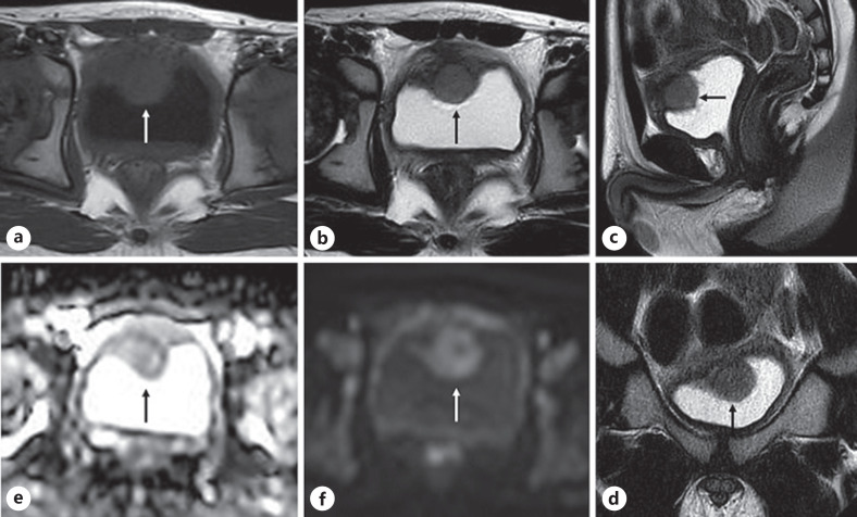
Fig. 2.
Pelvic MRI demonstrated the 18 × 17 × 16 mm broad-based mass (arrow), suggesting submucosal tumor in the dome of the bladder. The mass showed low-to-moderate signal intensity on T1-weighted images (a) and slight high signal intensity on T2-weighted images (b–d) and restricted diffusion with low signal intensity on apparent diffusion coefficient map (e) and abnormal high signal intensity on diffusion-weighed imaging (f).

