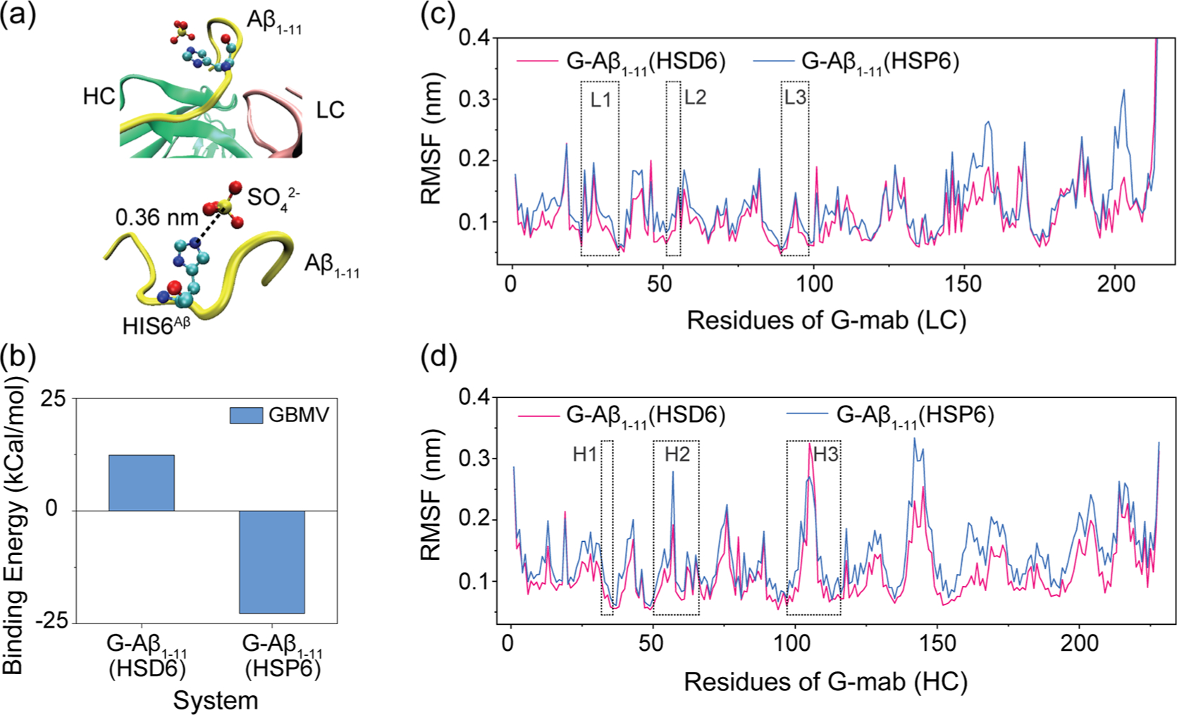
Figure 1.

Effects of protonation of His6Aβ on G-mab–Aβ1–11 complex. (a) Part of the G-mab–Aβ1–11 complex in crystal structure (PDB ID 5CSZ). Aβ1–11, light chain, and heavy chain are colored yellow, pink, and green, respectively. His6Aβ and SO42− are shown in VDW representation. A SO42− is located near the nitrogen atoms of His6Aβ side chain (the distance between NE2 and S is 0.36 nm), indicating that His6Aβ is protonated. (b) The binding energy between G-mab and Aβ1–11 with neutral His6 (HSD6) or protonated His6 (HSP6) in our MD simulations. (c, d) Cα root mean square fluctuation (Cα-RMSF) of G-mab’s light chain (c) and heavy chain (d). CDR loops are highlighted in boxes.
