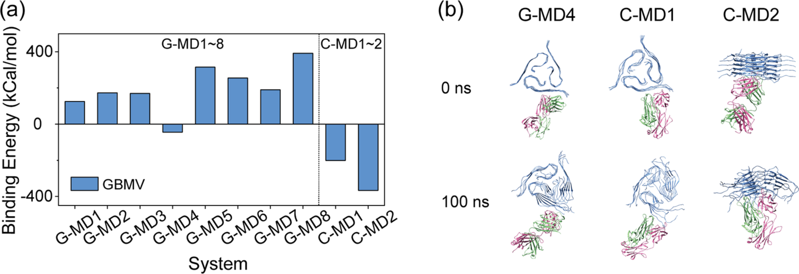
Figure 3.

Stabilities of the 10 antibody–Aβ4016mer structures obtained from docking. The initial and final structures of all the systems can be seen in Figure SI. (a) Average binding energy between Aβ40 fibril and antibodies. The binding energy is calculated by 〈Ebind〉 = 〈Ecomplex〉 − 〈Eantibody〉 – 〈Epeptide〉. (b) Initial and final structures of three antibody–Aβ40l6mer complexes with negative binding energy (C-MD1, C-MD2, and G-MD4). Aβ, HC, and LC are colored in blue, green, and pink, respectively.
